Frenectomy
What is it?
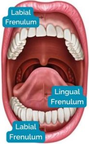
The frenulum is a muscular insertion located in different parts of the mouth that connect it to other moving structures, such as the lip or the tongue.
Where are they located?
- In the midline known as Medial Frenulae:
- Upper labial frenulum
- Lower labial frenulum
- Lingual frenulum
- Between the premolars known as Lateral Frenulae:
- Upper
- Lower
- Accessory
What should be consider when performing a frenectomy or frenulotomy
- Shape of the frenulum
- Where the frenulum is inserted according to Placek Mirko et al.
- Mucous (insertion at the mucogingival junction
- Gingival (insertion into attached gum)
- Papillary (insertion into the inter-incisor papilla)
- Papillary Penetrating (insertion into the inter-incisor papilla but penetrating to the palatine papilla. This is usually what causes diastemas between the central incisors.
- Composition of the frenulum
- Fibrous (composed of connective tissue and mucous membrane)
- Muscular (different muscles may be integrated within the frenulum)
- Mixed (a firm tendinous attachment to the floor of the mouth and a fibrous cord attached to the alveolar process)
Knowing these things we can make a diagnosis and be able to know:
- If necessary remove it
- What type of incisions are we going to make with the laser?
- What power are we going to use on the laser to be able to remove it?
The good news is that in many cases we do not have to remove them .
Medial frenulums with insertion into the mucosa are found in 69%
Medial frenulums with insertion into the gum are found in 31%
What do we know about lingual frenulums?
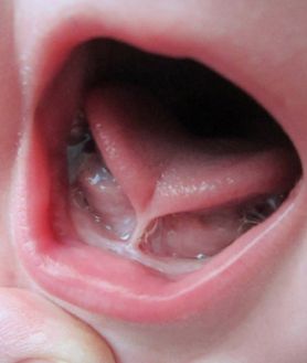
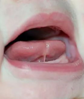
With respect to lingual frenulums, we must know that they are subdivided into 4 types:
1. Frenulum inserts on the tip of the tongue causing difficulties in speech
2. Frenulum inserts a few mm further back from the tip of the tongue allowing better movement of the tongue
3. Frenulum inserts on the base of the tongue (submucosal component)
4. There is no visible lingual frenulum
Pathologies associated with aberrant frenulums
Frenums are not considered pathologies of the oral cavity, but what we call aberrant frenums do cause changes in the protective periodontium and are considered mucogingival deformities and have a great impact on the patient’s function and aesthetics.
The braces that are inserted into:
1. The papilla
2. Those that penetrate the vestibular papillae to the palatine papilla
3. and the lingual ones
are most associated with:
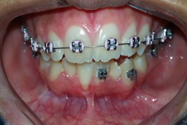
1. Gingival Recessions
On the lower teeth, we can observe gum recession. This permanent retraction ends in major periodontal disorders that can even lead to the loss of the lower central incisors.
2. Loss of papillae
3. Accumulation of bacterial plaque by preventing easy hygiene with the brush
4. Limitations of movement of the upper lip
In the upper jaw, we can see patients who have short smiles, in which the gums are very visible. This may be due to the presence of this type of frenulum and, by removing the frenulum, we obtain a noticeable improvement in the drooping of the lip without the need for gingival trimming or the placement of aesthetic veneers.
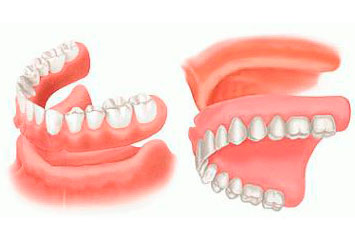
5. They can alter the retention and stability of removable prostheses , many times the prostheses do not seat properly due to the braces or they limit the extension of the prosthesis, altering its retention and stability.
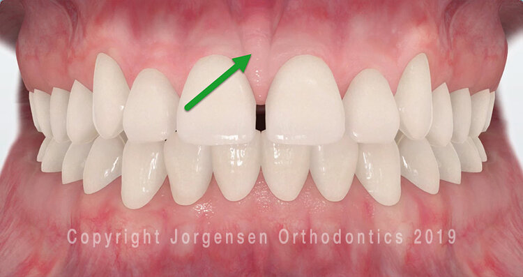
6. Diastemas in the upper incisors: In orthodontic and maxillary orthopedic treatments we often find diastemas caused by the insertion of these frenulums, which often have an insertion in the palate and, if not eliminated, make the orthodontic treatment not be effective.
The hypertrophic upper labial frenulum can cause a space between the central incisors that is larger than usual, giving rise to what we know as inter-incisal diastema.
How we remove the frenulum if necessary
Many dental professionals (general dentists, orthodontists) do not like to perform these frenectomies and refer them to periodontists who use diode or Erbium laser technology, since the results are magnificent. With this technology, less anesthesia is administered, recovery is faster and the procedures are less invasive. The diode laser causes less tissue damage, less bleeding, and does not require sutures.
In general, the patient’s body responds better to minimally invasive laser dentistry than to traditional procedures (anesthesia, cutting and suturing).


